IIT Madras has launched ANCHOR, a publicly accessible 3D human brainstem atlas at cell resolution, offering detailed maps for neuroscience research.
Chennai, June 12: The Indian Institute of Technology Madras (IIT Madras) has released what it describes as the world’s most detailed three-dimensional atlas of the human brainstem at cell resolution, marking a significant step in neuroscience research and brain mapping.
Developed by the Sudha Gopalakrishnan Brain Centre (SGBC), the platform, named ANCHOR (Atlas of Neurochemical Characterization of the Human Brainstem with 3D Reconstruction), provides comprehensive multi-modal maps of the human brainstem spanning prenatal, childhood and adult stages.
According to IIT Madras, the atlas contains detailed reconstructions of more than 200 brainstem nuclei and fibre tracts created from hundreds of serial tissue sections. Researchers overlaid eight complementary immunostains across more than 500 sections to identify and map distinct neurochemical cell types.
The institute has made ANCHOR publicly accessible through its dedicated portal to support researchers, clinicians and patients worldwide. The project forms part of SGBC’s broader effort to create cell-resolution maps of the human brain across different stages of life and disease conditions.
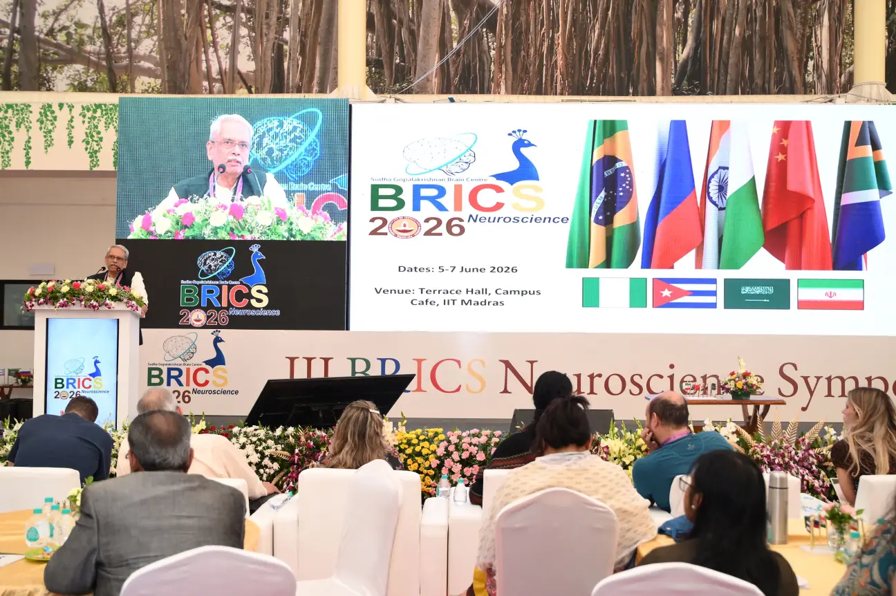
The atlas was formally released during the third BRICS Neuroscience Symposium 2026, held at the IIT Madras campus from June 5 to 7. The event brought together neuroscientists, clinicians, researchers and academicians from BRICS countries.
The release took place in the presence of Principal Scientific Adviser to the Government of India Prof. Ajay Kumar Sood, Infosys co-founder Kris Gopalakrishnan, IIT Madras Director Prof. V. Kamakoti, SGBC Head Prof. Mohanasankar Sivaprakasam and other researchers, industry representatives and supporters of the centre.
Addressing the event virtually, Prof. Ajay Kumar Sood described the atlas as a significant accomplishment in neurobiology. He said the framework integrates MRI imaging, histology and detailed chemo-architectural information into a single digital platform. According to him, the atlas could help identify specific cell populations affected by brainstem lesions, potentially aiding future clinical applications.
Prof. Sood also noted that the achievement follows the release of DHARANI by SGBC last year. He said the centre had emerged through collaboration among government agencies, industry partners, philanthropic organisations and researchers from multiple institutions and countries. He further acknowledged contributions from medical institutions including CMC Vellore, Kilpauk Medical College, MediScan Systems and Shri Ramachandra Institute of Higher Education and Research in obtaining brain samples of different ages for the project.
Highlighting the broader significance of the work, IIT Madras Director Prof. V. Kamakoti said the centre’s research places the institute at the forefront of efforts to understand the human brain. He noted that SGBC is also studying brains affected by conditions such as rabies, dementia and Alzheimer’s disease to better understand structural changes associated with neurological disorders.
Kris Gopalakrishnan, who has supported the centre’s activities, said making the atlas publicly available would help advance neuroscience research globally. He emphasised the importance of accessibility, affordability and inclusiveness in scientific and technological development.
Explaining the technological foundation behind ANCHOR, Prof. Mohanasankar Sivaprakasam said the atlas is powered by a multi-modal image visualisation framework that links large-scale MRI data with microscopic cellular images. The system allows researchers to move seamlessly from overall brain structures to cellular-level details while maintaining precise spatial correspondence between datasets.
He said the release represents an important scientific milestone for the centre as it works toward imaging more than 100 whole human brains across different ages and neurological diseases.
Independent experts attending the symposium also welcomed the initiative. Prof. Shubha Tole of the Tata Institute of Fundamental Research described the programme as a major interdisciplinary effort combining engineering, neuroscience and medicine. She said the atlas reflects a level of integration that is both unprecedented and scientifically valuable.
Speaking virtually, Prof. Mu-Ming Poo of the Chinese Academy of Sciences said the decision to begin with a brainstem atlas was important because the brainstem serves as the critical link between the brain and spinal cord and regulates essential functions such as breathing, sleep, wakefulness and movement. He also praised the collaboration between neuroscientists, clinicians, engineers and computing experts that made the project possible.
SGBC currently brings together more than 200 researchers, engineers and technicians and collaborates with around 20 international partners. The centre aims to build one of the most comprehensive collections of human brain maps at cellular resolution, covering both healthy development and neurological diseases.